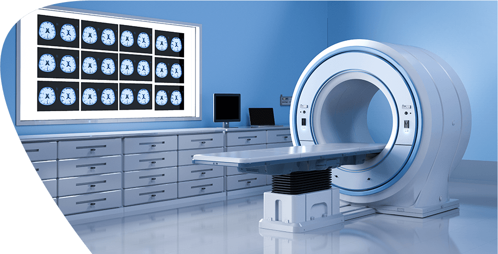
Imaging & Interventional Radiology
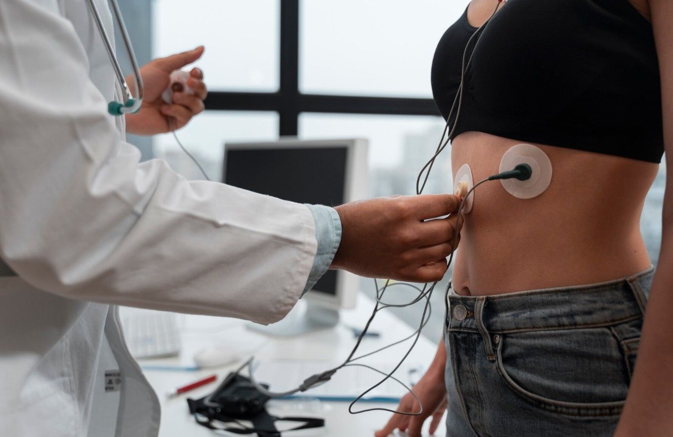
Interventional radiologists identify and manage various diseases. They work by using small tools like catheters or wires, which they insert into the body from the outside. To guide these procedures, they use imaging techniques such as X-rays, CT scans, and ultrasounds. Interventional radiology provides an alternative to surgery in many situations. It can sometimes even remove the need to stay in a hospital.
Why Should You Choose KIMSHEALTH for Interventional Radiology?
- The Radiology Department at KIMSHEALTH holds NABH MIS accreditation and offers top-notch imaging services with modern tools and advanced technology. The department operates a fully digital workflow supported by RIS and PACS systems.
- Diagnostic and Interventional Radiology services stay open around the clock every day.
- The center uses cutting-edge 3 Tesla MRI machines with Artificial Intelligence technology to speed up scan processes.
- A team of skilled Radiologists handles everything from basic imaging to advanced techniques across different areas. These include Neuro Radiology, Cardiac and Vascular Imaging, Pulmonary Radiology, Abdominal Imaging, Women’s Imaging, Paediatric Radiology, Musculoskeletal Radiology, Onco Imaging and Transplant Imaging.
- To train future specialists, the facility runs residency programs in Radiology and Interventional Radiology — DNB (Radiology) and DrNB (Interventional Radiology) — certified by the National Board of Examinations in New Delhi.
- Case discussions and clinico-radiological exchanges help to encourage ongoing learning and build sharper diagnostic skills. Senior consultants mentor younger radiologists to improve their expertise.
- Radiologists contribute through publishing papers and writing peer reviews in both national and international journals.
- They take part as faculty in state, national and global radiology conferences.
Treatments and Procedures Available
Radiologists and their teams handle advanced diagnostic imaging studies and carry out many types of image-aided therapies.
a. Radiography
- Digital Radiography
- Techniques like Fusion Imaging/stitch imaging
- Special View Radiography
- Digital Portable Radiography systems
b. Digital Fluoroscopy
- Barium Study for GIT
- Special contrast imaging for the Genitourinary system like IVU, RGU, MCU, and HSG
- Imaging procedures like Sinusograms and Fistulograms
- Exam of the tear ducts through Dacryocystography
- Salivary gland imaging using Sialography
- ENT Fluoroscopy
c. Digital Mammography with 3D Tomo Synthesis
d. Bone Density Scan / DEXA
e. Dental Radiography
- Orthopantomography (OPG)
- Lateral Cephalometric Radiograph (LCR)
- IOPA (Intra Oral Peri Apical)
- TMJ (Temporomandibular Joint Imaging)
- CBCT (Cone Beam Computed Tomography)
- ‘C’ Arm Image Intensifier Guided Procedures
- Procedures in Ortho, Neuro, Gastro, and Uro Surgeries using ‘C’ Arm guidance
- Endoscopy procedures performed with ‘C’ Arm imaging
- USG (Ultra Sono Graphy)
- Regular Ultrasonography
- Obstetric and Gynec Ultrasonography including Anomaly & NT Scans
- Colour Doppler Flow Imaging (CDFI)
- Mobile Ultrasonography
f. 128 Slice and 16 Slice Wide Bore CT (Computed tomography)
- Basic CT Scans with Plain and Contrast Imaging.
- CT Angiography to analyse the Brain, Pulmonary, Coronary, Liver, Renal, Aortic & limbs.
- Detailed 3D imaging using CT technology.
- Virtual procedures like Colonoscopy and Bronchoscopy.
- Robotic TKR Planning
- CT Simulation techniques.
g. MRI (Magnetic Resonance Imaging)
- 1.5 Tesla MRI
- Functional MRI (fMRI) - Advanced Brain Mapping
- Standard MRI scans of the full body with Plain or Contrast Imaging including Heart and Breast.
- Advanced MRCP to examine the bile ducts.
- MR Angiography both with and without contrast imaging.
- MRI Venography using contrast and without contrast
h. Advanced 3T MRI Studies
- Magnetic Resonance Spectroscopy
- Tractography
- Cardiac MRI scanning
- Measuring liver fat
- Cartilage imaging
- Full-body diffusion imaging
i. PET Scans (Positron Emission Tomography)
- FDG scans
- Gallium imaging
- PSMA scans
Diagnostic and Treatment Procedures:
- Brain and Neuro Vascular Interventions
- Abdominal and Hepatobiliary procedures
- Vascular access and line placement
- Aortic and Peripheral Vascular procedures
- DSA (Digital Subtraction Angiography)
- Interventional procedures done with Ultrasound or CT Guidance such as Biopsy, FNAC, catheter drainage, Fiducial marker implants and hook wire placements.
- PTBD (Percutaneous transhepatic biliary drainage)
- Biliary stenting
- TIPPS (Transjugular intrahepatic portosystemic shunt)
- Tumour embolisation techniques like TACE (Transarterial Chemoembolisation) and TARE (Transarterial radioembolisation)
- Ganglion blocks
- Ablation techniques including RFA (Radiofrequency Ablation) and MWA (Microwave Ablation)
Facilities and Equipment Provided
Important tools and machines available in the department:
- Functional MRI (fMRI) - Advanced Brain Mapping
- Fixed Digital Radiography: 3
- Mobile Digital Radiography: 3
- Digital Mammography using Tomo Synthesis: 1
- BMD or DEXA Scanner: 1
- OPG machine: 1
- IOPA devices: 7
- CBCT system: 1
- C-arm Image Intensifiers: 4
- High-end Ultrasound along with Colour Doppler Scanners: 16
- Mobile Ultrasound Scanners: 15
- CT Scanners (128-Slice and 16-Slice): 2
- MRI Scanners (3 Tesla and 1.5 Tesla): 2
- PET-CT Machine: 1
- Biplane Cathlab: 1
Interventional Radiology
A talk on Stroke and Intervention In stroke by Dr. Manish Kumar Yadav - Consultant and Coordinator, Imaging & Interventional Radiology, KIMSHEALTH Trivandrum
Our Doctors


Patient Testimonials

Ellea Jane Collister - Maternity
Ellea Jane Collister shares her experience of the most significant moment of her...
Obstetrics & Gynecology


Our Blogs
Electrophysiology
Heart rhythm abnormalities can be of two types-Tachycardia and bradycardia.Tachycardia or abnormal increase in heart rate can be physiological or pathological. This pathological increase can be easily identified from a surface ECG by an Electro physiologist. It is caused due to short circuits in the heart. The medical treatment of such tachycardias are often difficult and may be lifelong. More so, many tachycardia recur on medications. Electro physiological study involves the identification of such short circuits using special pacing techniques. After identifying the circuit and its location, it can be disconnected by using radio frequency waves, which is referred to as Radio frequency ablation. This method has been time tested and we are re-introducing this novel technique in KIMS. This technique offers the advantage that the patient can be off long term medications and the side effects of these medications. Moe over, in patients involved in certain kinds of jobs like that of pilot, Drivers, army personnel etc may be disqualified from their position due to such lethal arrhythmias. Electro physiological study and ablation can be of great help in such a situation. In the bradycardia sector electro physiology contributes immensely by the implantaion of pacemakers. The more advanced devices like impantable cardioverter defibrillatior (ICD ), has saved several lives by an appropriate shock. Such shocks can be life saving in patients with palpitations and syncope (unexplained loss of conciousness), with lethal arrhythmias. In patients with refractory heart failure also electo physiology has an edge over drugs.Cardiac re-synchronisation therapy (CRT) is yet another ray of hope in the horizon of patients with refractory heart failure. This procedure involves pacing the three chambers of the heart and “re -synchronising” the heart, thereby reducing the symptoms of heart failure.